| Clone ID | DM53 |
|---|---|
| Target | |
| Immunogen | PME100012 |
| Synonyms | B7-H3; CD276; B7 homolog 3; B7H3 |
| Host Species | Rabbit |
| Description | Biotinylated Anti-B7-H3 antibody(DM53); Rabbit mAb |
| Delivery | In Stock |
| Uniprot ID | Q5ZPR3 |
| IgG type | Rabbit IgG |
| Clonality | Monoclonal |
| Reactivity | Human |
| Applications | Flow Cyt |
| Recommended Dilutions | Flow Cyt 1:1000 |
| Purification | Purified from cell culture supernatant by affinity chromatography |
| Endotoxin | Less than 1.0 EU/μg by the LAL method. For <1 EU/mg requirements, please contact us for customization. |
| Formulation & Reconstitution | Lyophilized from sterile PBS, pH 7.4. Normally 5 % – 8% trehalose is added as protectants before lyophilization. Please see Certificate of Analysis for specific instructions of reconstitution. |
| Storage&Shipping | Store at -20°C to -80°C for 12 months in lyophilized form. After reconstitution, if not intended for use within a month, aliquot and store at -80°C (Avoid repeated freezing and thawing). Lyophilized proteins are shipped at ambient temperature. |
| Sterility | Products are supplied non-sterile. For cell culture applications, dilute in appropriate medium and sterile-filter (0.22 µm) prior to use. |
| Background | The protein encoded by this gene belongs to the immunoglobulin superfamily; and thought to participate in the regulation of T-cell-mediated immune response. Studies show that while the transcript of this gene is ubiquitously expressed in normal tissues and solid tumors; the protein is preferentially expressed only in tumor tissues. Additionally; it was observed that the 3' UTR of this transcript contains a target site for miR29 microRNA; and there is an inverse correlation between the expression of this protein and miR29 levels; suggesting regulation of expression of this gene product by miR29. Alternatively spliced transcript variants encoding different isoforms have been found for this gene. |
| Usage | Research use only |
| Conjugate | Biotinylated |
| DIMA Disclaimer | All DIMA recombinant antibodies are genuinely generated by DIMA Biotech. They are all under patent application. Any protein sequencing or reverse engineering attempt is prohibited. We are actively scr |
Biotinylated Anti-B7-H3 antibody(DM53); Rabbit mAb
Price: 10μg $139.00 ; 100 μg $670.00 ; 500 μg $1999.00
Product Data Dima FAQ
Images Dima FAQ

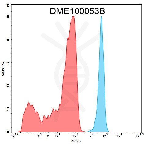
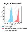
Figure 1. 1e5 of HEK293 cell line were stained with 100 μL of 1:1000 diluted Biotinylated Anti-B7-H3 antibody (DM53), Rabbit mAb (Blue histogram) or isotype control (Red histogram) respectively, washed and then stained with Streptavidin APC. The experimental samples were analyzed by flow cytometry.
Related Products

Biosimilar reference antibodies
SKU: BME100730 Target: B7-H3
Application: N/A
Price: 50μg $82.00 ; 100 μg $162.00
Biosimilar reference antibodies
SKU: BME100181P Target: B7-H3
Application: Flow Cyt
Price: 100 test $192.00
Biosimilar reference antibodies
SKU: BME100010 Target: B7-H3
Application: ELISA; Flow Cyt
Price: 50μg $82.00 ; 100 μg $162.00
Overexpression Stable Cell Line
SKU: CEL100101 Target: B7-H3
Application: FACS Data
Price:Inquiry
ECD Proteins
SKU: PME100012 Target: B7-H3 Tag: C-Mouse Fc and 6×His Tag
Expression Host: HEK293
Price: 10μg $82.00; 50μg $320.00 ; 100 μg $480.00