| Clone ID | DM77 |
|---|---|
| Target | |
| Immunogen | PME100039 |
| Synonyms | CD33;SIGLEC3;gp67 |
| Host Species | Rabbit |
| Description | Anti-CD33 antibody(DM77); Rabbit mAb |
| Delivery | In Stock |
| Uniprot ID | P20138 |
| IgG type | Rabbit IgG |
| Clonality | Monoclonal |
| Reactivity | Human |
| Applications | ELISA; Flow Cyt |
| Recommended Dilutions | ELISA 1:5000-10000; Flow Cyt 1:100 |
| Purification | Purified from cell culture supernatant by affinity chromatography |
| Endotoxin | Less than 1.0 EU/μg by the LAL method. For <1 EU/mg requirements, please contact us for customization. |
| Formulation & Reconstitution | Lyophilized from sterile PBS, pH 7.4. Normally 5 % – 8% trehalose is added as protectants before lyophilization. Please see Certificate of Analysis for specific instructions of reconstitution. |
| Storage&Shipping | Store at -20°C to -80°C for 12 months in lyophilized form. After reconstitution, if not intended for use within a month, aliquot and store at -80°C (Avoid repeated freezing and thawing). Lyophilized proteins are shipped at ambient temperature. |
| Sterility | Products are supplied non-sterile. For cell culture applications, dilute in appropriate medium and sterile-filter (0.22 µm) prior to use. |
| Background | Sialic-acid-binding immunoglobulin-like lectin (Siglec) that plays a role in mediating cell-cell interactions and in maintaining immune cells in a resting state. Preferentially recognizes and binds alpha-2,3- and more avidly alpha-2,6-linked sialic acid-bearing glycans. Upon engagement of ligands such as C1q or syalylated glycoproteins; two immunoreceptor tyrosine-based inhibitory motifs (ITIMs) located in CD33 cytoplasmic tail are phosphorylated by Src-like kinases such as LCK. These phosphorylations provide docking sites for the recruitment and activation of protein-tyrosine phosphatases PTPN6:SHP-1 and PTPN11:SHP-2. In turn; these phosphatases regulate downstream pathways through dephosphorylation of signaling molecules. One of the repressive effect of CD33 on monocyte activation requires phosphoinositide 3-kinase:PI3K. |
| Usage | Research use only |
| Conjugate | Unconjugated |
| DIMA Disclaimer | All DIMA recombinant antibodies are genuinely generated by DIMA Biotech. They are all under patent application. Any protein sequencing or reverse engineering attempt is prohibited. We are actively scr |
Anti-CD33 antibody(DM77); Rabbit mAb
Price: 10μg $99.00 ; 100 μg $446.00 ; 500 μg $1340.00
Product Data Dima FAQ
Images Dima FAQ
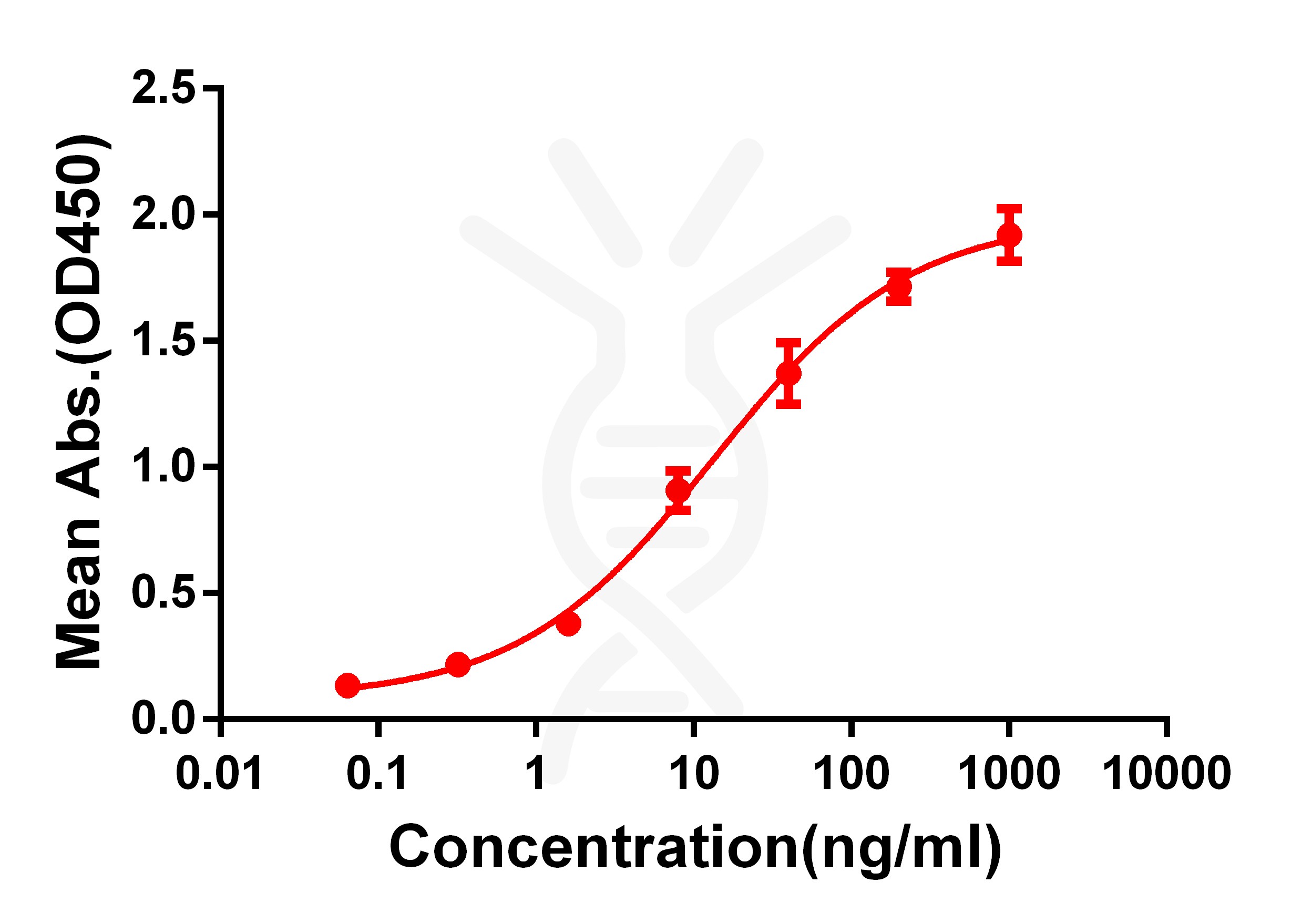
Figure 1. ELISA plate pre-coated by 2 μg/ml (100 μl/well) Human CD33 protein, hFc-His tagged protein PME100039 can bind Rabbit anti-CD33 monoclonal antibody (clone: DM77) in a linear range of 1-100 ng/ml.
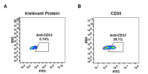
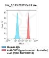
Figure 2. HEK293 cell line transfected with irrelevant protein (A) and human CD33 (B) were surface stained with Rabbit anti-CD33 monoclonal antibody 1μg/ml (clone: DM77) followed by Alexa 488-conjugated anti-rabbit IgG secondary antibody.
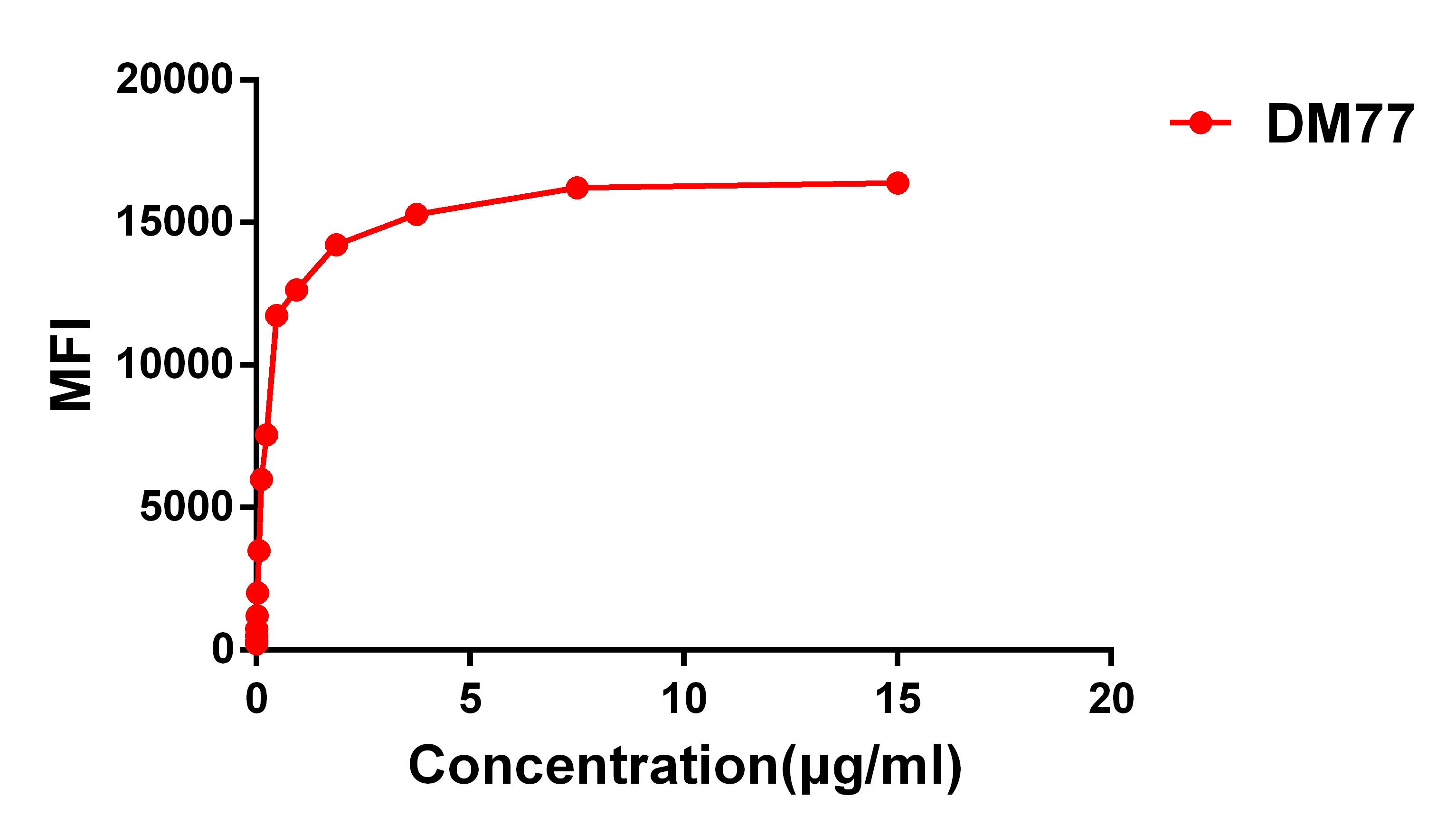
Figure 3. Flow cytometry data of serially titrated Rabbit anti-CD33 monoclonal antibody (clone: DM77) on HEK293 cell line transfected with human CD33. The Y-axis represents the mean fluorescence intensity (MFI) while the X-axis represents the concentration of IgG used.
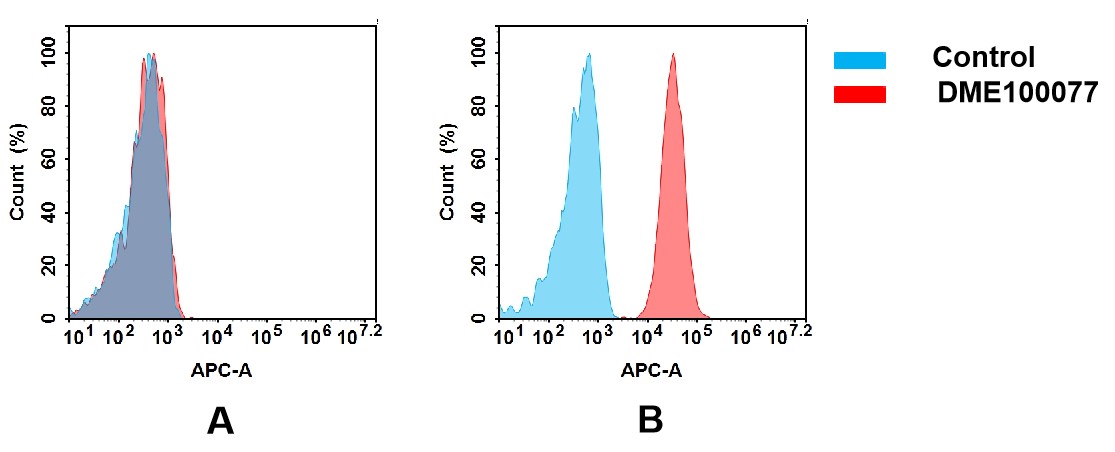
Figure 4. Flow cytometry analysis of antigen binding of rabbit anti-human CD33 mAb(DME100077).
(A) DME100077 does not bind to 293T cells that do not express CD33.
(B) A clear peak shift of DME100077 was seen compared to the control when incubated with CD33-expressing THP-1 cells, indicating strong binding of DME100077 to CD33. Antibodies were incubated at 10 μg/mL.
Related Products
ECD Proteins
SKU: PME-M100015 Target: CD33 Tag: C-Human Fc Tag
Expression Host: HEK293
Price: 10μg $72.00; 50μg $274.00 ; 100 μg $411.00
Monoclonal antibodies
SKU: DME100077B Target: CD33
Application: ELISA; Flow Cyt
Price: 10μg $139.00 ; 100 μg $670.00 ; 500 μg $1999.00
Overexpression Stable Cell Line
SKU: CEL100025 Target: CD33
Application: FACS Data
Price:Inquiry
ECD Proteins
SKU: PME100693 Target: CD33 Tag: C-Human Fc Tag
Expression Host: HEK293
Price: 10μg $72.00; 50μg $272.00; 100μg $409.00

Biosimilar reference antibodies
SKU: BME100419 Target: CD33
Application: N/A
Price: 50μg $82.00 ; 100 μg $162.00