
Aquaporins (AQPs) are best known as membrane channels that transport water. As research has advanced, however, their biological significance has expanded far beyond basic water transport to include cerebral edema, autoimmunity, brain fluid clearance, and neurodegenerative diseases. Among the AQP family, aquaporin-4 (AQP4) has attracted particular attention because of its distinctive distribution and complex regulation in the central nervous system (CNS).
A 2026 study explored targeted delivery of the AQP4 inhibitor TGN-020 to ischemic stroke lesions, with the aim of reducing cerebral edema while preserving glymphatic function as much as possible [1]. The findings highlight an important feature of AQP4 as a drug target: therapeutic benefit cannot be reduced to simple channel inhibition, but instead depends on disease stage, spatial localization, and functional state. So what makes AQP4 structurally and mechanistically unique, and why is it emerging as a potential therapeutic target?
1. AQP4 and the Aquaporin Family
Aquaporins are transmembrane channel proteins widely expressed in cell membranes. They primarily facilitate the rapid movement of water, and in some cases small solutes, along osmotic gradients. The mammalian aquaporin family generally comprises 13 members, AQP0-AQP12, with broad tissue distribution across the kidneys, nervous system, lungs, liver, gastrointestinal tract, eyes, skin, and adipose tissue. Based on sequence features and permeability, AQPs can be broadly divided into three groups: classical aquaporins, including AQP0, AQP1, AQP2, AQP4, AQP5, AQP6, and AQP8, which mainly transport water; aquaglyceroporins, including AQP3, AQP7, AQP9, and AQP10, which can also transport glycerol, urea, and other small solutes; and superaquaporins/unorthodox aquaporins, including AQP11 and AQP12, which are more commonly associated with intracellular membrane systems and have distinct structural and functional properties [2].
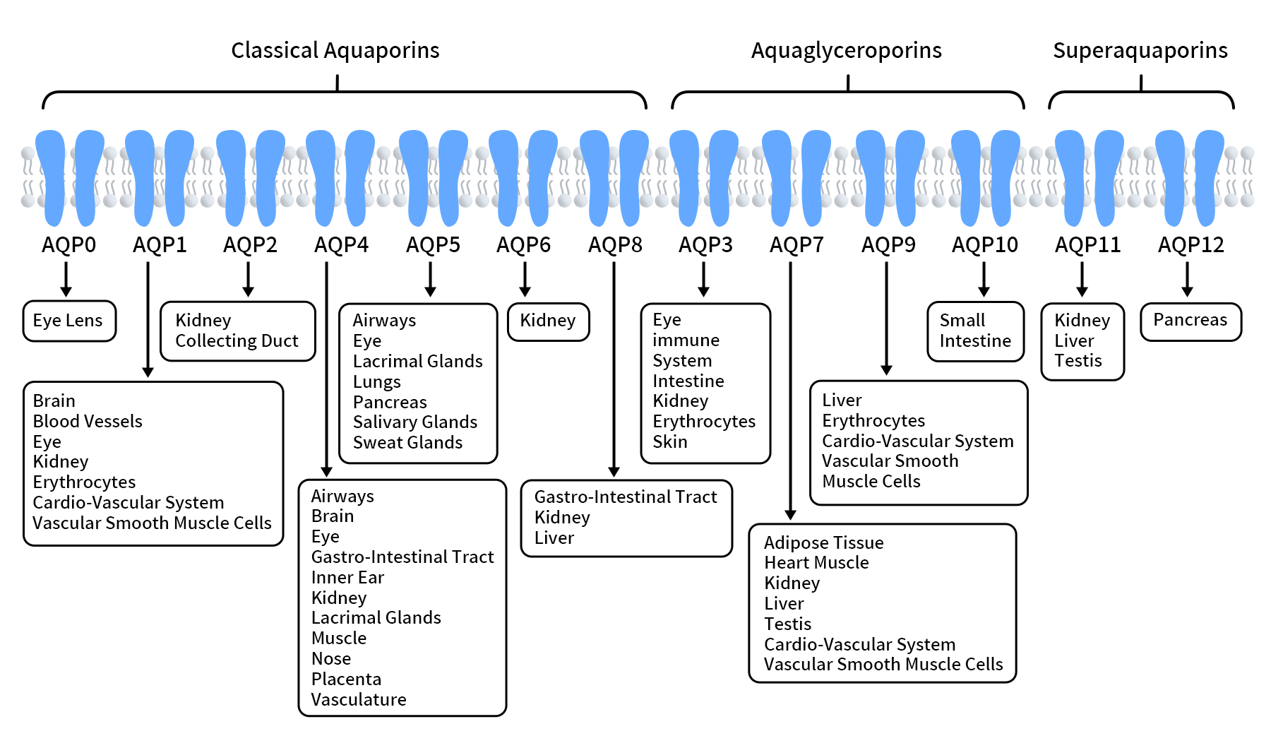
Figure 1. Three Classes of Aquaporins [3]
Individual AQP family members show marked tissue specificity. AQP0 is mainly found in the lens, AQP2 is a key regulator of water reabsorption in the renal collecting duct, and AQP5 contributes to fluid secretion in the salivary glands, lacrimal glands, and respiratory tract. AQP4, by contrast, is one of the most abundant and functionally important water channels in the CNS. It is enriched in astrocytes, particularly at perivascular astrocyte endfeet [2,4]. This distinctive localization places AQP4 at the intersection of brain water homeostasis, the blood-brain barrier, the glymphatic system, and multiple neurological diseases.
2. Structure of AQP4
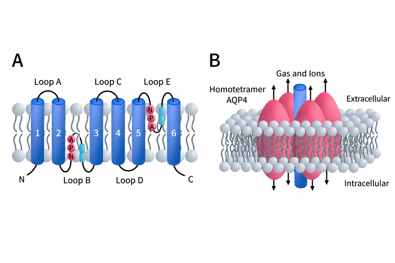
Like most aquaporins, an AQP4 monomer has a characteristic six-pass transmembrane α-helical architecture connected by five loops (Loops A-E). Loops B and E fold inward from opposite sides of the membrane and contain the highly conserved NPA motifs (Asn-Pro-Ala). These two NPA-containing regions approach each other near the center of the membrane and contribute to formation of the selective water-conducting pore [2].
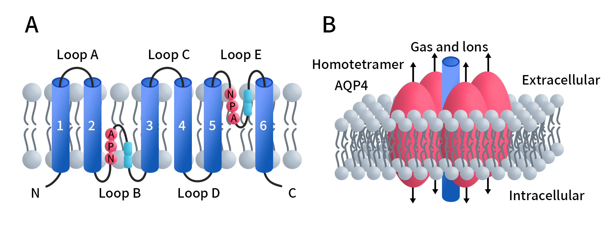
Figure 2. Structure of AQP4 [2]
Multiple AQP4 isoforms have been described, including AQP4a, AQP4b, AQP4c, AQP4d, AQP4e, AQP4f, and AQP4-Δ4. The best-characterized isoforms are AQP4a (M1) and AQP4c (M23). Both retain the full six-pass transmembrane architecture, but M23 lacks the first 22 N-terminal amino acids present in M1. This relatively small difference has a major effect on higher-order AQP4 assembly.
AQP4 monomers first assemble into tetramers, and multiple tetramers can further organize into orthogonal arrays of particles (OAPs). M23 has a strong intrinsic tendency to form OAPs, whereas M1 generally co-assembles with M23 and limits further expansion of the array. As a result, the M1/M23 ratio helps determine OAP size and organization. In the CNS, OAPs are particularly enriched at perivascular astrocyte endfeet. Thus, the native state of AQP4 is shaped not only by its transmembrane architecture, but also by isoform composition, tetramerization, and higher-order OAP assembly [5].
A 2025 cryo-EM study reconstituted full-length AQP4 M1 in lipid nanodiscs and resolved its tetrameric structure at 2.1 Å resolution. The study further showed that higher-order AQP4 assembly can influence the presentation of extracellular conformational epitopes [6]. This is important for antibody discovery and drug screening: an intact amino acid sequence alone may not be sufficient, because the way AQP4 assembles within a membrane environment can also affect target recognition.
3. Distribution, Core Functions, and Mechanism of AQP4
AQP4 is primarily expressed in astrocytes in the CNS and is highly enriched at perivascular astrocyte endfeet, the ependymal lining, and brain-cerebrospinal fluid interfaces [4,7]. This polarized distribution places AQP4 at key sites of exchange among blood, cerebrospinal fluid (CSF), and interstitial fluid (ISF), where it contributes to brain water homeostasis, cell-volume regulation, and the formation and clearance of cerebral edema.
Mechanistically, AQP4 does not consume adenosine triphosphate (ATP) or actively pump water. Instead, it functions as a bidirectional water channel that rapidly facilitates water movement according to osmotic gradients across the plasma membrane. The high local density of AQP4 at astrocyte endfeet can enhance water exchange and contributes to CSF-ISF exchange and glymphatic-associated fluid transport [2,4].
AQP4 function also depends on whether the protein is localized to the right place. Its membrane localization and polarization are regulated at multiple levels, including cytoskeletal anchoring, isoform assembly, and post-translational modification. When this polarized distribution is disrupted, water transport and brain fluid-clearance functions may be impaired even without a major reduction in total AQP4 expression [7].
4. AQP4 in Neurological Diseases
4.1 AQP4 and Cerebral Edema
The role of AQP4 in cerebral edema is strongly stage-dependent. During the early cytotoxic edema phase of ischemic stroke, disruption of ion homeostasis drives water into astrocytes, and AQP4 can accelerate this influx. Later, when blood-brain barrier disruption and vasogenic edema predominate, AQP4 may instead facilitate clearance of excess water from brain tissue into the blood or cerebrospinal fluid. AQP4 can therefore have opposing effects at different stages of edema, underscoring why drug development must consider the timing of intervention rather than relying on prolonged, indiscriminate AQP4 inhibition [4].
4.2 AQP4 and NMOSD
In neuromyelitis optica spectrum disorder (NMOSD), AQP4 shifts from being a water channel to serving as a major autoantigen. AQP4-IgG recognizes AQP4 on the surface of astrocytes and can trigger complement- and cell-mediated immune injury, leading to astrocyte damage followed by demyelination and neuronal injury [6,8]. This mechanism gives AQP4 dual relevance as both a disease biomarker and a therapeutic target. Unlike cerebral edema strategies that directly modulate channel activity, NMOSD-directed approaches focus on blocking the interaction between pathogenic AQP4-IgG and native conformational epitopes, or on suppressing downstream immune effector mechanisms.
4.3 AQP4, the Glymphatic System, and Neurodegenerative Diseases
In the glymphatic system, the key issue is not simply how much AQP4 is expressed, but how strongly it is polarized to perivascular astrocyte endfeet. Loss of AQP4 polarization may reduce CSF-ISF exchange and metabolic waste clearance and has been associated with sleep disorders, brain aging, and multiple neurodegenerative diseases [7].
In Alzheimer’s disease (AD), abnormal AQP4 polarization has been linked to impaired clearance of β-amyloid (Aβ), tau, and other metabolites. However, this area is still driven largely by mechanistic and animal studies, and the causal relationship between impaired brain-fluid clearance and human disease progression remains to be established. AQP4 is therefore better viewed as an emerging therapeutic target in AD rather than one that has already been clinically validated.
5. Advances in AQP4-Targeted Drug Development
Direct AQP4-targeted drug development remains at an early translational stage, and no therapy that directly targets AQP4 has yet been approved. Among publicly disclosed programs, AER-271/SIM0800 has completed Phase I studies and is one of the most advanced direct AQP4 small-molecule programs, whereas TGN-020, ORI-TRN-002, TGN-073, and Aquaporumab remain primarily at the preclinical stage. The field is also moving beyond simple channel blockade toward a broader set of strategies, including channel inhibition, functional enhancement, modulation of AQP4 polarization, and blockade of pathogenic autoantibody binding.
5.1 Small-Molecule AQP4 Inhibitors
Small-molecule inhibitors are currently the most advanced class of direct AQP4-targeted agents. Their primary rationale is to reduce AQP4-mediated water influx during the early phase of ischemia and thereby mitigate cytotoxic edema.
AER-271 is the most representative clinical-stage program in this category. AER-271 is an intravenous prodrug of the active AQP4 inhibitor AER-270. It was originally developed by Aeromics and was subsequently advanced by Simcere in Greater China. Phase I studies have been completed in both the United States and China; to date, publicly available information has not shown that a Phase II trial has formally started.
TGN-020 has long been used as a classical research tool for AQP4 inhibition. In 2026, a lesion-targeted delivery strategy was used to enrich TGN-020 in ischemic stroke regions, with the aim of reducing cerebral edema while minimizing disruption of whole-brain glymphatic function [1]. In addition, Origenis reported the novel AQP4 inhibitor ORI-TRN-002 in 2024. In Xenopus oocytes expressing AQP4, ORI-TRN-002 showed an IC50 of 2.9 ± 0.6 μM and remains in preclinical optimization [9].
5.2 AQP4 Enhancers
In contrast to inhibition strategies, AQP4 enhancers aim to increase channel function or restore perivascular localization, with a particular focus on glymphatic function and neurodegenerative disease research.
TGN-073 is a representative AQP4 facilitator/enhancer. A 2026 study in PS19 tauopathy mice reported that TGN-073 enhanced glymphatic-associated CSF influx, reduced tau accumulation, neuronal loss, and glial responses, and restored perivascular AQP4 enrichment. These effects were largely lost in AQP4-deficient mice, supporting an AQP4-dependent mechanism [10].
A similar concept has appeared in early drug-discovery efforts. In 2025, Shandong First Medical University disclosed an AQP4-related purine compound, Compound 61, reported to bind AQP4, enhance AQP4 polarization in cellular assays, and reduce astrocyte senescence. The program remains at an early discovery stage.
5.3 Antibody and Antigen-Specific Immunotherapy Strategies
Another distinctive AQP4-directed strategy arises from NMOSD. Here, the goal is not to change water permeability, but to prevent pathogenic AQP4-IgG from binding AQP4 on the astrocyte surface.
Aquaporumab is a representative engineered anti-AQP4 monoclonal antibody developed by researchers at the University of California, San Francisco (UCSF). Its Fc region was engineered to eliminate complement-dependent cytotoxicity (CDC) and antibody-dependent cellular cytotoxicity (ADCC) while preserving high-affinity AQP4 binding. It can therefore competitively occupy extracellular AQP4 epitopes and block binding of pathogenic patient AQP4-IgG. Aquaporumab remains at the preclinical stage [11].
Together, these approaches highlight a central feature of AQP4 pharmacology: the therapeutic objective is not simply to turn the channel “off” or “on,” but to modulate AQP4 in a disease-, stage-, and localization-dependent manner.
6. DIMA BIOTECH’s Nanodiscs: Supporting Native AQP4 Research and Drug Discovery
The structural and disease-related findings discussed above point to the same challenge: pharmacological recognition of AQP4 depends strongly on correct folding and higher-order assembly in a native-like membrane environment. In NMOSD, for example, antibody recognition involves extracellular conformational epitopes, and the 2025 cryo-EM study used full-length AQP4 reconstituted in lipid nanodiscs for structural analysis [6]. For antibody screening and binding studies, therefore, access to full-length AQP4 in a native-like state can be more important than simply having the protein sequence available.
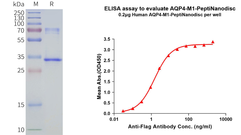
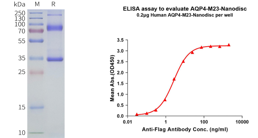
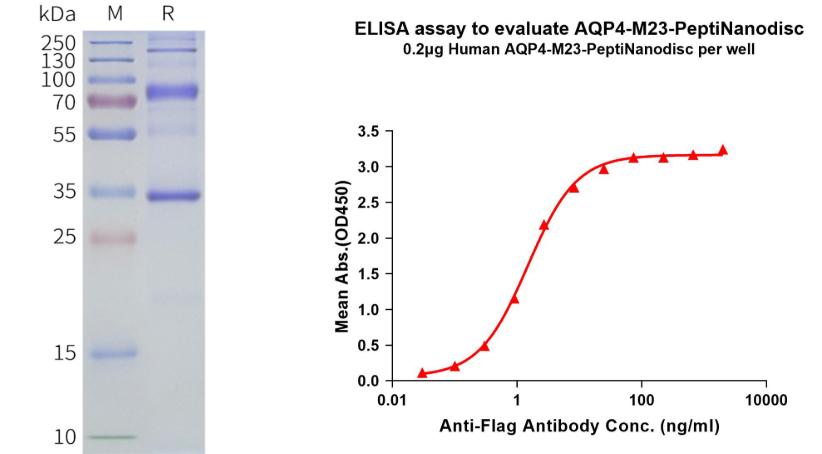
To address the challenge of preserving native conformations of multi-pass membrane proteins, DIMA BIOTECH has developed full-length AQP4 proteins in two Nanodisc formats using its proprietary membrane protein expression platform: polymer-based and peptide-based. Polymer-based Nanodiscs use polymers to stabilize AQP4 and its surrounding membrane-like environment, helping preserve the intact transmembrane architecture and native-like conformation. Peptide-based Nanodiscs use a peptide stabilization system and contain no phospholipids, making them particularly suitable for cell-based assays and applications where phospholipid background may interfere. Both formats retain full-length AQP4 and can support antibody discovery and screening, conformational antibody-binding studies, cell-based assays, drug-binding evaluation, and mechanistic studies of AQP4.
Human AQP4-M1 full-length protein - synthetic nanodisc ( FLP100504)
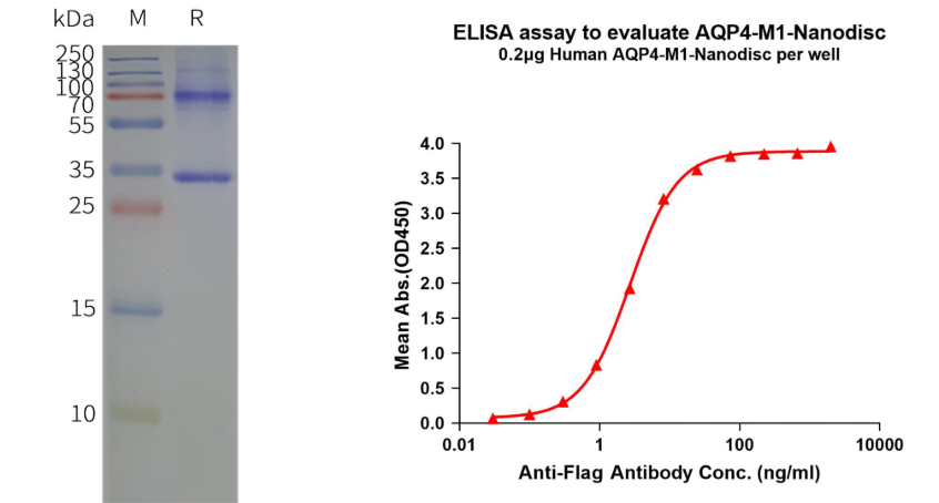
Human AQP4-M1 full-length protein - PeptiNanodisc (FLP400504)
Human AQP4-M23 full-length protein - synthetic nanodisc (FLP100509)
Human AQP4-M23 full-length protein - PeptiNanodisc (FLP400509)
Related Products
References
- Jin L, Yang Z, Wei B, et al. Amelioration of Post-Stroke Edema and Microcirculatory Dysfunction via Targeted AQP4 Inhibition While Preserving the Glymphatic System. Advanced Science. 2026;13(10):e20118.
- Li Y, Wang Y, Huang X, et al. Role of aquaporins in brain water transport and edema. Frontiers in Neuroscience. 2025;19:1518967.
- Dajani S., Saripalli A., Sharma-Walia N. Water transport proteins–aquaporins (AQPs) in cancer biology. Oncotarget. 2018; 9: 36392-36405.
- García Ríos CA, Leon-Rojas JE. Aquaporin-4 in Stroke and Brain Edema—Friend or Foe? International Journal of Molecular Sciences. 2025;26(17):8178.
- Potokar M, Jorgačevski J, Zorec R. Astrocyte Aquaporin Dynamics in Health and Disease. Int J Mol Sci. 2016;17(7):1121.
- Gupta M, Khandelwal NK, Nelson A, et al. Structural basis of aquaporin-4 autoantibody binding in neuromyelitis optica. Science Advances. 2025;11(8):eadq7560.
- Ma D, Zhang H. Astrocytic aquaporin-4 expression, localization, and polarization: mechanisms and therapeutic potential in neurological diseases. Neuroscience. 2026;592:52–62.
- Holian AH, Weinshenker BG. Emerging Role of Targeted Monoclonal Antibodies in Neuromyelitis Optica Spectrum Disorders. BioDrugs. 2025;39:573–589.
- Thormann M, Traube N, Yehia N, et al. Toward New AQP4 Inhibitors: ORI-TRN-002. Int J Mol Sci. 2024;25(2):924.
- Yamada K, Ishida K, Sakamoto A, et al. AQP4-dependent enhancement of glymphatic function attenuates tau pathology and neurodegeneration in PS19 mice. Mol Neurodegener. 2026;21:43.
- Duan T, Tradtrantip L, Phuan PW, et al. Affinity-matured ‘aquaporumab’ anti-aquaporin-4 antibody for therapy of seropositive neuromyelitis optica spectrum disorders. Neuropharmacology. 2020;162:107827.
