
Kir7.1, an inwardly rectifying potassium channel encoded by the KCNJ13 gene, is a member of the Kir channel family. In 2026, Kir7.1 research advanced rapidly in both structural biology and precision therapeutic development. Multi-state Cryo-EM structures revealed how phosphatidylinositol 4,5-bisphosphate (PIP₂), cholesterol, and bioactive steroids modulate channel conformation; prime editing of the KCNJ13 L144P variant restored channel function; and anticodon-engineered transfer RNA (ACE-tRNA) promoted targeted readthrough of the W53X premature termination codon and improved electroretinographic responses in Leber congenital amaurosis type 16 (LCA16) mouse model [1–3]. Additional structural studies clarified the molecular basis of the R162W loss-of-function phenotype and the pore-blocking mechanism of ML418 [5,10]. Together, these advances position Kir7.1 at the intersection of retinal ion homeostasis, channel structural biology, and precision therapy.
1. Kir7.1 and the Inwardly Rectifying Potassium Channel Family
Members of the Kir family share a common two-transmembrane architecture but differ substantially in tissue distribution, gating mechanisms, and physiological roles. Kir2.x channels are strong inward rectifiers that make major contributions to resting membrane potential; Kir3.x channels, also known as G protein-gated inwardly rectifying potassium (GIRK) channels, are regulated primarily by GPCR signaling; and Kir6.x channels assemble with sulfonylurea receptor (SUR) subunits to form ATP-sensitive potassium (KATP) channels that couple cellular metabolism to membrane excitability [12,14]. Kir1.1 (encoded by KCNJ1), GIRK1/Kir3.1 (encoded by KCNJ3), Kir2.3 (encoded by KCNJ4), and Kir7.1 illustrate the functional diversification of Kir channels across renal ion transport, GPCR signaling, membrane-potential control, and retinal homeostasis.
Kir7.1 is a highly divergent member of the Kir family with distinctive biophysical and pharmacological properties. Compared with many other Kir channels, it has an exceptionally low unitary conductance and weak inward rectification. Its macroscopic conductance also shows an unusual relationship with extracellular K⁺ concentration and can increase as extracellular K⁺ falls under RPE-relevant conditions [12–14]. These properties are well suited to its role in epithelial ion transport, particularly in the retinal pigment epithelium (RPE).
2. Structure of Kir7.1
Kir7.1 has the canonical Kir-channel topology, with two transmembrane α-helices (M1 and M2) in each subunit. M1 is followed by an extracellular turret region and an intramembrane pore-forming segment that includes the pore helix and the selectivity filter (¹¹⁹TIGYGYT¹²⁴), before reconnecting to M2. Both the N- and C-termini face the cytoplasm, and four Kir7.1 subunits assemble into a functional homotetramer around a central ion-conduction pore [1,5]. Cryo-EM studies published in 2026 resolved key gating features, including the inner helix gate at the cytoplasmic end of M2, with Val157 contributing to the constriction, and the cytoplasmic G-loop gate, whose narrow point is centered near Ile281 [1]. Kir7.1 also contains the distinctive Met125 residue adjacent to the selectivity filter. In many other Kir channels, the equivalent position is occupied by a positively charged arginine; substitution by Met125 prevents the canonical salt-bridge interaction with Glu115 and creates a more permissive outer-filter environment, which likely contributes to the unusual Cs⁺ permeation properties of Kir7.1 [1]. The disease-associated L114P variant lies within the pore-forming region and markedly impairs channel function; it has been linked to autosomal-dominant snowflake vitreoretinal degeneration (SVD) with retinoschisis [11].
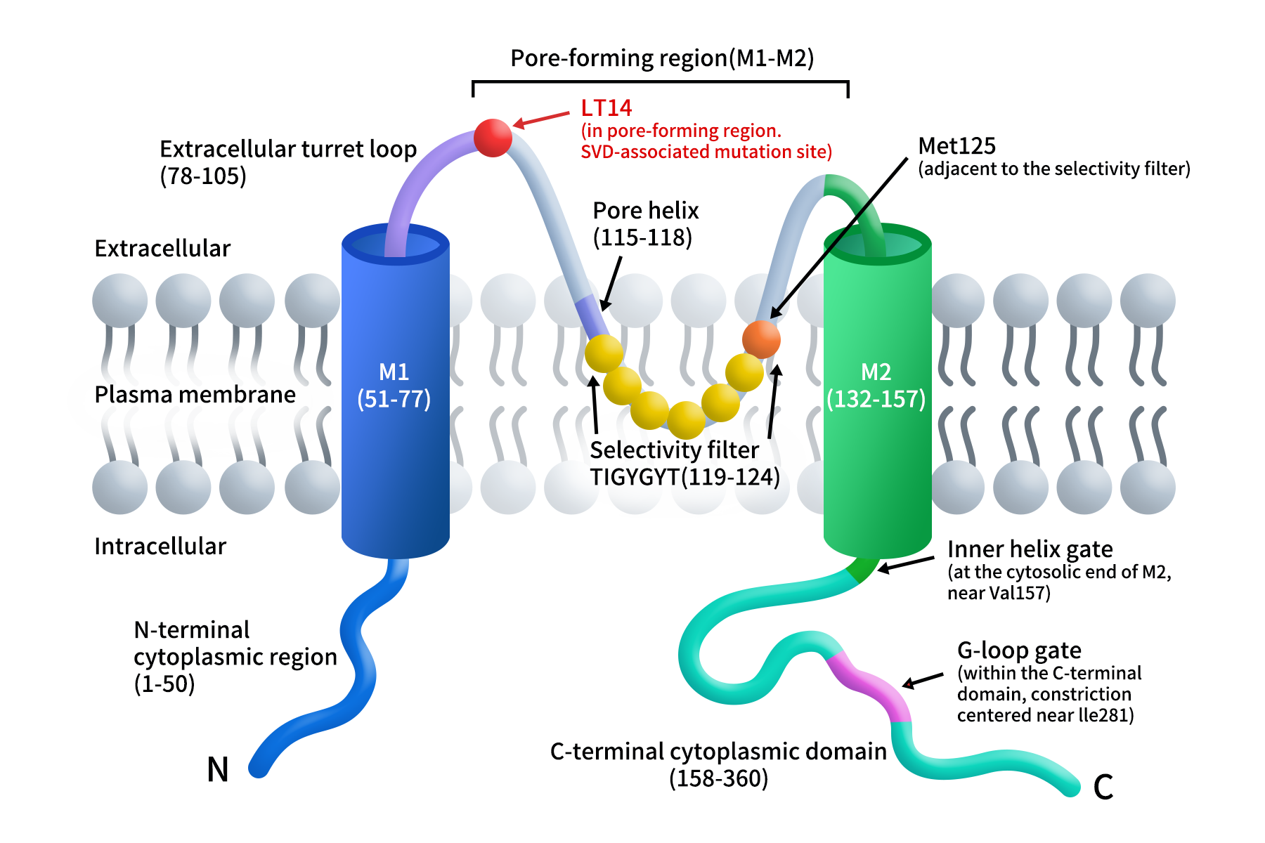
Figure 1. Schematic topology of a human Kir7.1 subunit.
These structural findings provide a direct framework for interpreting disease-associated variants and small-molecule modulation. The 2026 R162W cryo-EM structure showed that the bulky tryptophan side chain reorients toward the central pore, creating a steric obstruction to K⁺ permeation and providing a structural explanation for the dominant-negative loss-of-function phenotype [10]. In a separate study, ML418 was resolved in complex with the R162Q variant, where it occupies the central cavity below the selectivity filter and is associated with rearrangements of both the transmembrane and cytoplasmic domains, consistent with stabilization of a closed or inactivated-like conformation [5].
3. Tissue Distribution and Physiological Functions of Kir7.1
Kir7.1 is expressed in several tissues, including the RPE, brain, small intestine, thyroid, and other epithelia, but its role in the RPE is the best characterized. In native human RPE, Kir7.1 is highly enriched at the apical membrane and contributes to subretinal K⁺ homeostasis, transepithelial ion and fluid transport, and maintenance of membrane potential [4,12,13]. Patient-derived and pluripotent-stem-cell-derived RPE models further show that Kir7.1 dysfunction is associated with impaired phagocytosis of photoreceptor outer segments and disruption of normal RPE organization, highlighting a broader role in RPE integrity and photoreceptor support [13].
In the central nervous system, Kir7.1 has also been implicated in signaling associated with the hypothalamic melanocortin-4 receptor (MC4R), a key regulator of energy balance and feeding behavior. A 2026 study provided additional evidence for close physical and functional coupling between Kir7.1 and MC4R at the plasma membrane, while also noting that the possible contribution of intermediary signaling components has not been fully excluded [5].
4. Mechanisms of Kir7.1 Function
In the RPE, a key physiological role of Kir7.1 is to help buffer changes in K⁺ concentration within the subretinal space.
Following illumination, cyclic nucleotide-gated (CNG) channels in photoreceptor outer segments close, and the K⁺ concentration in the subretinal space falls from approximately 5 mM to about 2 mM. Kir7.1 has the unusual property that its macroscopic K⁺ conductance can increase as extracellular K⁺ decreases, helping the apical RPE membrane respond to light-induced changes in the subretinal ionic environment. Together with Na⁺/K⁺-ATPase and the Na⁺-K⁺-2Cl⁻ cotransporter (NKCC), Kir7.1 participates in apical K⁺ recycling and contributes to transepithelial ion and fluid transport [4,12].
Kir7.1 gating is also modulated by membrane lipids and steroids. The 2026 multi-state cryo-EM study identified endogenous PIP₂ at a conserved cytoplasmic binding site and showed that membrane lipids are linked to large conformational rearrangements of the cytoplasmic domain. Cholesterol was associated with a less active channel conformation, whereas progesterone and dehydroepiandrosterone (DHEA) enhanced Kir7.1 activity. PIP₂ potentiated progesterone-mediated activation but was not strictly required for steroid-induced activation under the experimental conditions used [1].
In the nervous system, a G protein-independent mode of MC4R–Kir7.1 coupling has been proposed. Earlier work showed that α-melanocyte-stimulating hormone (α-MSH) inhibits Kir7.1 and depolarizes MC4R-expressing neurons, whereas agouti-related peptide (AgRP) promotes Kir7.1 opening and neuronal hyperpolarization [7]. In 2026, NanoBiT proximity assays and receptor–channel fusion constructs provided further support for a close physical and functional association between MC4R and Kir7.1, although the complete molecular mechanism remains unresolved [5].
5. Kir7.1 and Inherited Retinal Diseases
The best-established disease associations of Kir7.1 are inherited retinal degenerations, including LCA16, SVD, and some forms of retinitis pigmentosa (RP).
LCA16 is typically caused by biallelic loss-of-function variants in KCNJ13 and is inherited in an autosomal-recessive manner. Affected individuals may present in infancy or early childhood with severe visual impairment, nystagmus, and markedly abnormal electroretinography (ERG). Reduced or absent Kir7.1 function disrupts RPE ion homeostasis and other RPE cellular functions, ultimately compromising long-term photoreceptor integrity [4,13].
SVD, in contrast, is associated with dominant KCNJ13 variants. R162W is a well-established dominant-negative loss-of-function variant, and the L114P variant reported in 2026 also showed functional evidence consistent with a dominant-negative mechanism [10,11]. Structural analysis of R162W demonstrated a direct steric block of the K⁺ permeation pathway rather than a simple change in ion selectivity [10]. The adjacent R162Q variant has been associated with a milder, adult-onset form of RP and, in the 2026 structural and electrophysiological study, displayed a gain-of-function phenotype with a preference for a more open channel conformation [5].
6. Advances in Kir7.1 Precision Therapies and Drug Development
Therapeutic development around Kir7.1 currently follows two broad directions. For loss-of-function disorders such as LCA16, the goal is to restore Kir7.1 expression or activity through gene augmentation, genome editing, or premature termination codon (PTC) suppression. In parallel, direct Kir7.1 small-molecule inhibitors are being used primarily as pharmacological probes and as starting points for exploring indications beyond LCA16.
6.1 AAV-Mediated Gene Augmentation: HUB-101
HUB-101, developed by Hubble Therapeutics, is an AAV-based gene-augmentation program designed to deliver a functional KCNJ13 coding sequence to the RPE. Preclinical studies have demonstrated functional rescue in patient-derived human induced pluripotent stem cell-derived RPE (hiPSC-RPE) and animal models. Hubble Therapeutics reports that HUB-101 has received FDA Rare Pediatric Disease and Orphan Drug designations and has advanced into GMP-grade vector manufacturing and IND-enabling development. As of September 2026, publicly available pipeline information still lists HUB-101 as preclinical, and no registered human clinical trial was identified [15,16].
6.2 Genome Editing: From Base Editing to Prime Editing
Genome-editing strategies aim to correct pathogenic KCNJ13 variants at their endogenous locus. A 2023 study used SNCs to deliver ABE8e mRNA and a sgRNA, correcting the W53X variant and restoring Kir7.1 function in patient-derived hiPSC-RPE and an LCA16 mouse model [8]. In 2026, work on the L144P variant showed that cytosine base editors could achieve high editing efficiencies but generated bystander edits that limited functional rescue; prime editing achieved approximately 20% on-target correction without bystander nucleotide changes and restored Kir7.1 activity in 28% of analyzed cells [2]. A separate 2026 genome-wide CRISPR screen identified GJB2 and BET1L as cellular barriers to nonviral delivery: reducing either factor increased LNP-mediated base-editing efficiency by more than 3.5-fold in patient-derived RPE and enabled functional Kir7.1 recovery in a subset of edited cells [17].
6.3 Nonsense Suppression: From Small Molecules to Engineered tRNA
For nonsense variants such as W53X and R166X, PTC suppression offers a route to restore full-length Kir7.1 without directly rewriting the genomic sequence. Earlier aminoglycoside-derived readthrough compounds, including NB84 (also known as ELX-03) and ELX-01, showed functional rescue in W53X cellular models but did not progress into sustained LCA16 clinical programs. In 2026, ACE-tRNA provided a more codon-directed strategy: ACE-tRNA^Trp.UAG selectively recognized the UAG stop codon created by W53X, restored full-length membrane-localized Kir7.1 and channel activity in patient-derived hiPSC-RPE, and partially restored RPE function and ERG responses after subretinal delivery in an LCA16 mouse model [3].
6.4 Direct Kir7.1 Small-Molecule Inhibitors: Pharmacological Tools and Structure-Guided Research
Several small-molecule pore inhibitors have been developed as Kir7.1 pharmacological tools. VU590 was among the earliest examples; subsequent screening at Vanderbilt University identified VU714, which was optimized to yield the more selective inhibitor ML418 with an IC₅₀ of approximately 0.31 μM [9,19]. In 2026, cryo-EM analysis of the ML418-bound R162Q channel localized the compound to the central pore cavity beneath the selectivity filter and linked binding to a closed or inactivated-like conformation, providing a structural basis for future structure-guided ligand design [5]. Other screening hits, including MRT00200769, have further demonstrated the pharmacological tractability of Kir7.1, but these compounds remain research tools rather than clinical candidates for LCA16 [18].
7. DIMA Biotech Full-Length Kir7.1 Protein Supports Drug Discovery
Kir7.1 is a technically challenging membrane-protein target because its function depends on tetrameric assembly, coupling between the selectivity filter and gating regions, and the surrounding lipid environment [1,5]. Consequently, isolated linear peptides or protein fragments outside a membrane-like environment may not adequately reproduce conformational epitopes or ligand-binding states relevant to the native channel.
DIMA Biotech’s membrane-protein expression platform incorporates full-length Kir7.1 into a phospholipid bilayer using Nanodisc technology, providing a membrane-like format designed to preserve the intact transmembrane architecture and native-like conformational features of the protein in an aqueous system. Compared with short peptides or partial fragments, full-length Kir7.1 Nanodisc protein can provide a more physiologically relevant reagent format for conformational antibody screening, binding validation, ligand studies, and structure-guided drug discovery.
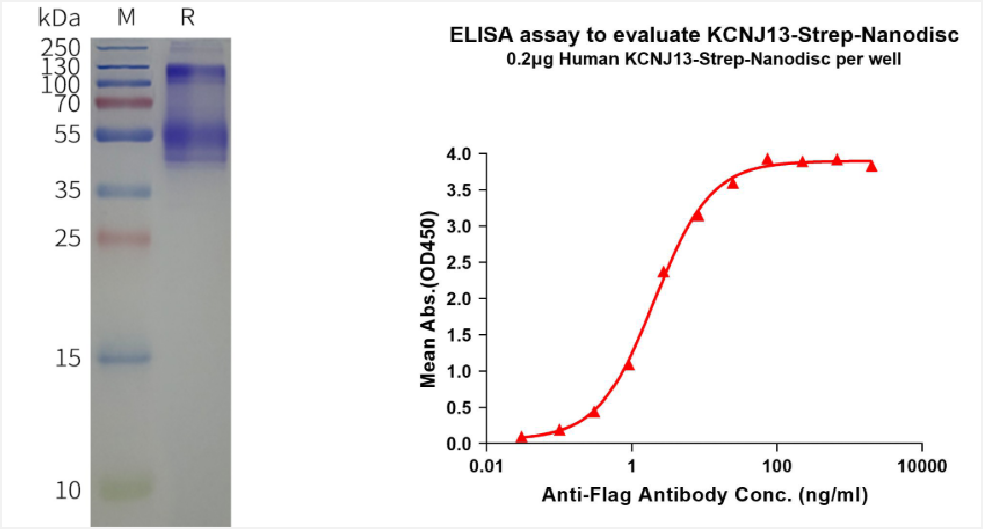
Human KCNJ13-Strep full length protein-synthetic nanodisc (FLP120520)
Related Products:
|
Target |
Cat.No. |
Product name |
|
KCNJ1 |
Human KCNJ1-Strep full length protein-synthetic nanodisc |
|
|
KCNJ3 |
Human KCNJ3-Strep full length protein-synthetic nanodisc |
|
|
KCNJ4 |
Human KCNJ4-Strep full length protein-synthetic nanodisc |
|
|
KCNJ13 |
Human KCNJ13-Strep full length protein-synthetic nanodisc |
References
- Niu Q, Vu S, Xu Y, et al. Bioactive lipid-mediated structural and functional regulation of the essential human potassium channel Kir7.1. Nature Communications. 2026;17:2764.
- Kabra M, Moosajee M, Navarrete A, et al. Synonymous editing alters ion channel function, favoring prime editing for retinal disease correction. International Journal of Biological Sciences. 2026;22(9):4670–4690.
- Shahi PK, Akyuz E, Gissot L, et al. Engineered tRNA reduces vision loss in a mouse model of Leber congenital amaurosis. Signal Transduction and Targeted Therapy. 2026;11:225.
- Kumar M, Pattnaik BR. Focus on Kir7.1: physiology and channelopathy. Channels (Austin). 2014;8(6):488–495.
- Peisley A, Hernandez CC, Dahir NS, et al. Structural and functional studies of inward rectifier Kir7.1 and its regulation by the Melanocortin-4 receptor. Nature Communications. 2026;17:9131.
- NCBI Gene. KCNJ13 potassium inwardly rectifying channel subfamily J member 13. Gene ID: 3769.
- Ghamari-Langroudi M, Digby GJ, Sebag JA, et al. G-protein-independent coupling of MC4R to Kir7.1 in hypothalamic neurons. Nature. 2015;520:94–98.
- Kabra M, Shahi PK, Wang Y, et al. Nonviral base editing of KCNJ13 mutation preserves vision in a model of inherited retinal channelopathy. Journal of Clinical Investigation. 2023;133(19):e171356.
- Raphemot R, Swale DR, Dadi PK, et al. ML418: The First Selective, Sub-Micromolar Pore Blocker of Kir7.1 Potassium Channels. ACS Chemical Neuroscience. 2016;7(7):1013–1023.
- O’Malley N, Nasrallah C, Churchill A, et al. Cryo-EM structure of the human Kir7.1 channel reveals the molecular basis of snowflake vitreoretinal degeneration disease. Communications Biology. 2026. doi:10.1038/s42003-026-10436-0.
- Pattnaik BR, Nischal KK, Alekseev O, et al. Association of Autosomal Dominant Snowflake Vitreoretinal Degeneration with Retinoschisis. Ophthalmology Science. 2026;6(6):101189.
- Beverley KM, Pattnaik BR. Inward rectifier potassium (Kir) channels in the retina: living our vision. American Journal of Physiology-Cell Physiology. 2022;323(3):C772–C782.
- Xu P, Zou W, Yin W, et al. Ion channels research in hPSC-RPE cells: bridging benchwork to clinical applications. Journal of Translational Medicine. 2024;22:1073.
- Hernandez CC, Gimenez LE, Dahir NS, et al. The unique structural characteristics of the Kir7.1 inward rectifier potassium channel: a novel player in energy homeostasis control. American Journal of Physiology-Cell Physiology. 2023;324(3):C694–C706.
- Hubble Therapeutics. HUB-101/LCA16 gene therapy program. Official program information, accessed September 2026.
- Hope in Focus. LCA Therapy Development Pipeline. Accessed September 2026.
- Saxena S, Kabra M, Abdeen AA, et al. Genome-wide CRISPR screening identifies cellular factors controlling nonviral genome editing efficiency. Nature Communications. 2026;17:8086.
- Wright PD, Kanumilli S, Tickle D, et al. A High-Throughput Electrophysiology Assay Identifies Inhibitors of the Inwardly Rectifying Potassium Channel Kir7.1. Journal of Biomolecular Screening. 2015;20(6):739–747.
- Lewis LM, Bhave G, Chauder BA, et al. High-Throughput Screening Reveals a Small-Molecule Inhibitor of the Renal Outer Medullary Potassium Channel and Kir7.1. Molecular Pharmacology. 2009;76(5):1094–1103.
