
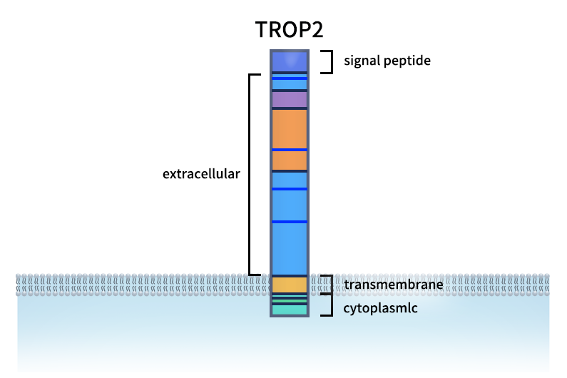
Trophoblast cell-surface antigen 2 (TROP2), also known as TACSTD2, EGP-1, and GA733-1, is a cell-surface glycoprotein encoded by the TACSTD2 gene and belongs to the tumor-associated antigen family. As an important tumor-associated target, TROP2 is involved in biological processes such as cell proliferation, calcium signaling, and tumor progression. Aberrant TROP2 expression has been associated with the development and progression of certain cancers and with unfavorable prognosis [1,2].
From a drug-development perspective, the extracellular domain of TROP2 can be recognized by specific antibodies. Following target binding, some anti-TROP2 antibodies can also be internalized into tumor cells, providing an opportunity to deliver cytotoxic payloads through ADCs. These characteristics have made TROP2 an important target in the development of ADCs for solid tumors.
In recent years, TROP2 has been clinically validated in several tumor types, including breast and lung cancer. Research has also expanded beyond conventional antibodies and ADCs to include bispecific antibodies, cell therapies, and other innovative therapeutic modalities. For an overview of representative agents and the latest clinical data, see: Development of TROP2-Targeted Therapeutics: ADC Clinical Pipeline and Future Directions.
1. TROP2 Protein Structure and Expression
TROP2 is a type I, single-pass transmembrane cell-surface glycoprotein consisting of 323 amino acids, with a theoretical molecular weight of approximately 35 kDa for the unmodified protein. Its structure can be divided into four main regions: a hydrophobic signal peptide (amino acids 1–26), which directs the protein into the secretory pathway; an extracellular domain (amino acids 27–274), which is the principal region recognized and bound by antibodies and contains a cysteine-rich region, a thyroglobulin type-1 domain, and several potential N-linked glycosylation sites (N33, N120, N168, and N208); a transmembrane domain (amino acids 275–297), which anchors TROP2 in the cell membrane; and a cytoplasmic tail (amino acids 298–323), which participates in intracellular signaling and contains a PIP2-binding-related region and phosphorylation sites [1–3]. Because of glycosylation, the apparent molecular weight of TROP2 expressed in mammalian cells may be higher than its theoretical molecular weight.
In addition, the TROP2 cytoplasmic tail contains several functional sites involved in signaling regulation. Ser303 is an important PKC-associated phosphorylation site. Studies have shown that mutation of Ser303 weakens the ability of TROP2 to promote tumor-cell growth. Structural studies further indicate that Ser303 phosphorylation can alter the charge distribution and conformation of the intracellular tail and affect the accessibility of key functional regions, suggesting that it may regulate TROP2 function through modulation of intracellular signaling [4].
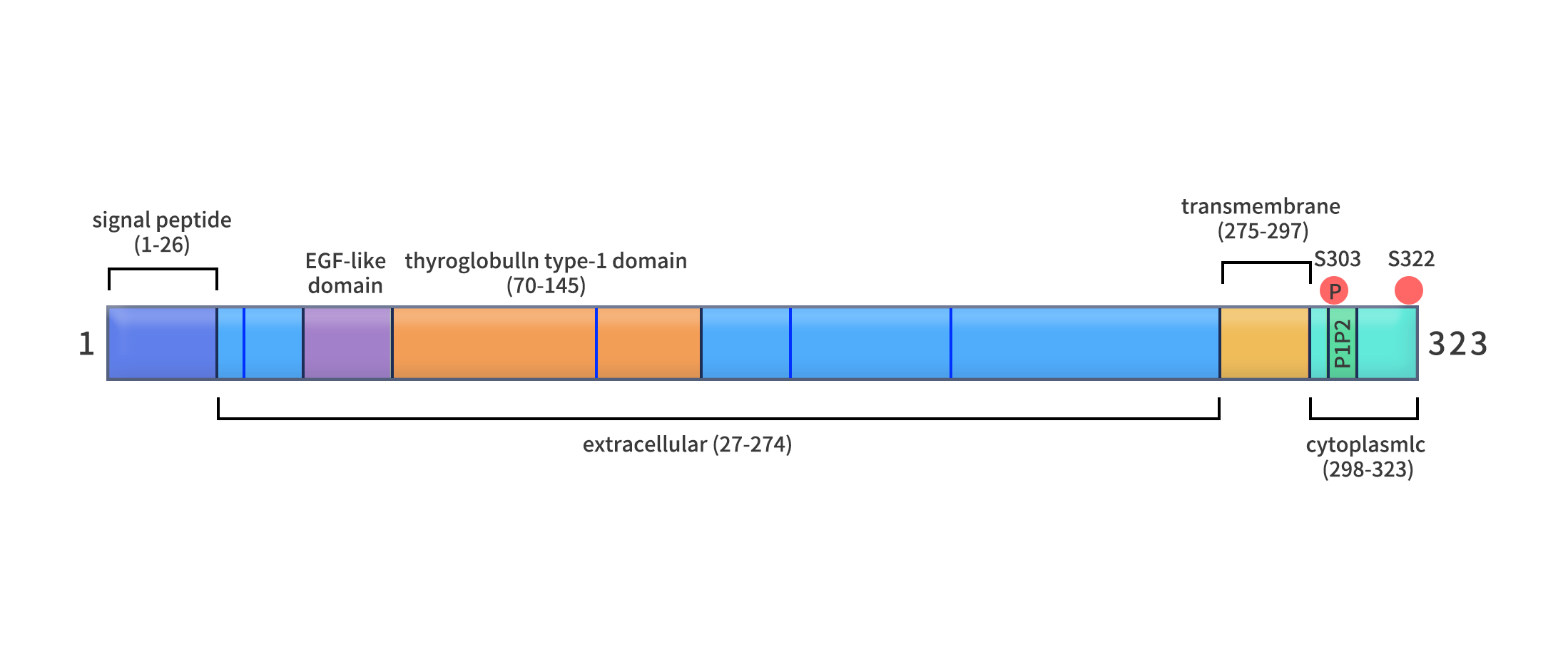
Figure 1. Structure of the TROP2 protein [1].
TROP2 is expressed at elevated levels, to varying degrees, in a range of epithelial-derived cancers, including breast, lung, gastric, colorectal, pancreatic, prostate, ovarian, and cervical cancers. However, TROP2 is not tumor-specific; it is also detectable in epithelial cells in certain normal tissues, including the skin, cervix, esophagus, salivary glands, breast, kidney, and respiratory tract. Its expression level, membrane localization, and intracellular distribution vary by tissue and cell type [1,5]. Therefore, differences in TROP2 expression between tumors and normal tissues, together with its accessibility on the cell surface, must both be considered when assessing its value as an ADC target and the potential for toxicity to normal tissues.
2. TROP2-Mediated Cellular Signaling Pathways
TROP2 is not merely a cell-surface antigen; it can also regulate multiple cellular signaling processes through phosphorylation of its cytoplasmic tail, lipid interactions, and protein complexes. Reported effects involve cell proliferation, survival, calcium signaling, cell-cycle regulation, adhesion, and migration. However, the direction of these effects depends on tumor type and cellular context, and not all of these pathways operate simultaneously in every model [2,5].
- PI3K/AKT, MAPK, and JAK2/STAT3 Signaling Pathways
In lung adenocarcinoma models, TROP2 has been reported to interact with IGF-1. The investigators proposed that TROP2 may bind and sequester IGF-1, reducing its effective interaction with IGF-1R and thereby attenuating IGF-1R activation and downstream signaling. Experimental findings showed that TROP2 overexpression reduced IGF-1R phosphorylation and suppressed AKT-related signaling and ERK activation, suggesting that TROP2 negatively regulates IGF-1R-associated signaling in this specific model [6]. By contrast, in glioblastoma models, TROP2 expression has been associated with activation of JAK2/STAT3 signaling, accompanied by increased cell proliferation and metastatic capacity [7]. Changes in cell-cycle and apoptosis regulators, including Cyclin D1, Cyclin E, CDKs, and Bcl-2/Bax, have also been reported [5,7].
- PIP2-Mediated Calcium Signaling and the MAPK/AP-1 Pathway
The TROP2 cytoplasmic tail contains a region associated with PIP2 binding. Studies have proposed that the interaction between TROP2 and PIP2 may affect the availability of PIP2 to phospholipase C (PLC). PLC hydrolyzes PIP2 to generate IP3 and DAG. IP3 can promote Ca²⁺ release from the endoplasmic reticulum, while DAG and Ca²⁺ contribute to PKC activation and may further influence MAPK signaling and the activity of the transcription factor AP-1. This signaling process has been linked to cell proliferation, epithelial–mesenchymal transition (EMT), and tumor angiogenesis [1,4,5].
- Regulation of Cell Adhesion and Motility
Unlike Ser303, Ser322 is another phosphorylation site mediated by PKCα/δ. In colorectal cancer cell models, Ser322 phosphorylation has been associated with reduced Claudin-7 protein levels, decreased localization at cell borders, and increased cell motility. These findings suggest that TROP2 can influence cell adhesion and motility through the PKCα/δ–Ser322–Claudin-7 axis [8]. In prostate cancer cell models, TROP2 can also affect FAK-related adhesion signaling and the interaction between cells and fibronectin through the RACK1/β1-integrin axis [9].
- β-Catenin-Related Signaling and Tumor Metastasis
In certain tumor models, binding of Galectin-3 to TROP2 can induce TROP2 phosphorylation and subsequent protein processing. The intracellular C-terminal fragment of TROP2 may participate in β-catenin/TCF4-associated transcriptional regulation and affect the expression of ZEB1 and E-cadherin. In addition, a gastric cancer study found that increased TROP2 expression was associated with downregulation of DSG2 and involved the EGFR/AKT and DSG2/PG/β-catenin signaling networks [5,10,11].
Taken together, TROP2-associated signaling involves multiple regulatory networks, including IGF-1R, JAK2/STAT3, PIP2/PLC/PKC, FAK, and β-catenin. Because the tumor types, cellular models, and experimental conditions differ across studies, these pathways should be understood as reported, context-dependent mechanisms rather than as a single fixed signaling cascade that occurs simultaneously in all tumors.
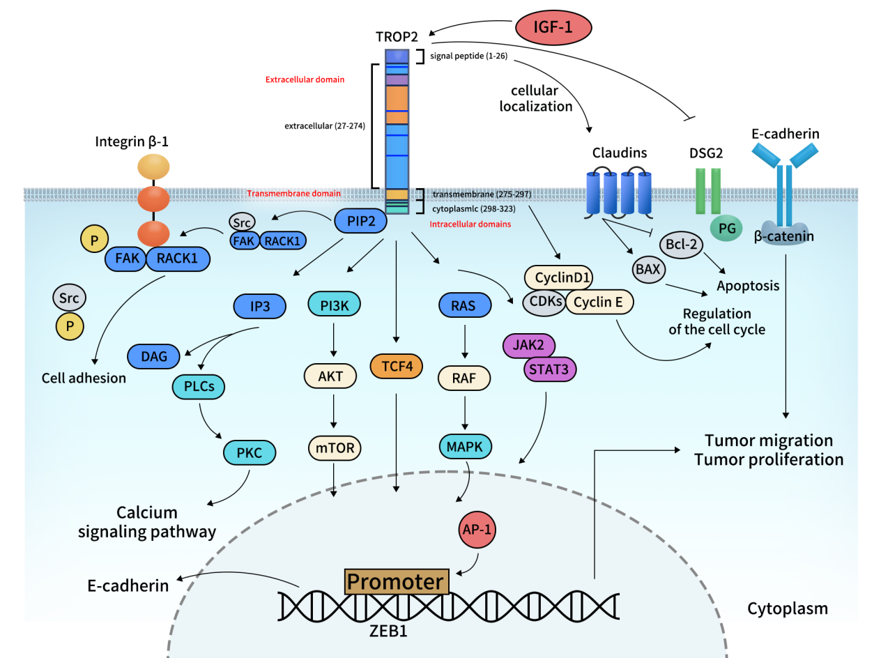
Figure 2. Proposed signaling mechanisms associated with TROP2 [5].
3. Roles of TROP2 in Tumor Development and Progression
Aberrant TROP2 expression and functional regulation are associated with multiple aspects of tumor biology. In some tumor models, TROP2 affects cell-cycle regulators such as Cyclin D1, Cyclin E, and CDKs, and is associated with anti-apoptotic changes such as increased Bcl-2 and decreased Bax, thereby promoting tumor-cell proliferation and survival [5,7,12]. These effects are dependent on tumor type and cellular context and should not be generalized to mean that TROP2 has the same pro-proliferative role in every cancer.
During tumor invasion and metastasis, TROP2 can alter cell adhesion and motility by affecting intercellular junctions and interactions between cells and the extracellular matrix. For example, PKCα/δ-mediated phosphorylation of Ser322 can affect the localization of Claudin-7, while regulation involving the RACK1/β1-integrin/FAK axis can alter tumor-cell adhesion to the extracellular matrix. In addition, some gastric cancer models have shown that increased TROP2 expression is accompanied by DSG2 downregulation and involves EGFR/AKT and DSG2/PG/β-catenin signaling [8–11].
Of particular interest, TROP2 protein processing itself may influence tumor progression. A study published in Neoplasia in 2021 found that ADAM10 cleaves the extracellular domain of TROP2 at the R87–T88 site. In colon cancer cell and animal models, this process was associated with tumor growth and metastasis, while mutation of the corresponding cleavage site weakened these effects [13]. This ADAM10-mediated extracellular cleavage should be distinguished from intramembrane proteolytic processes described in other studies that involve the release of the intracellular C-terminal fragment.
4. Therapeutic Development Potential of TROP2
The cell-surface expression of TROP2, the accessibility of its extracellular domain to antibodies, and the ability of some antibody-bound TROP2 molecules to undergo internalization provide a basis for antibody-based drug development. However, because TROP2 is also expressed in certain normal epithelial tissues, development of TROP2-targeted therapeutics must take into account differences in expression between tumor and normal tissues, membrane localization, internalization efficiency, and potential on-target toxicity.
Current drug-development efforts targeting TROP2 mainly fall into the following categories:
- Antibody therapeutics: Anti-TROP2 monoclonal antibodies can be used for target recognition, expression analysis, and related biological studies. Key considerations in their development include binding specificity, epitope recognition, affinity, and cell-surface binding capacity.
- Antibody-drug conjugates (ADCs): ADCs are currently the most clinically advanced therapeutic modality targeting TROP2. By conjugating an anti-TROP2 antibody to a cytotoxic payload, ADCs enable target-specific recognition and intracellular drug delivery. Representative agents include sacituzumab govitecan, datopotamab deruxtecan, and sacituzumab tirumotecan.
- Bispecific antibodies and other novel modalities: As TROP2 ADC development continues to advance, new therapeutic formats are also entering the research landscape, including dual-targeting ADCs, dual-payload ADCs, and TROP2-targeted CAR-T and CAR-NK cells.
These strategies are intended to expand the therapeutic potential of TROP2 targeting through multi-target recognition, combinations of different mechanisms of action, and recruitment of immune cells.
Development of TROP2-Targeted Therapeutics: ADC Clinical Pipeline and Future Directions.
5. DIMA Biotech’s TROP2 Products and Research Solutions
To support antibody discovery, screening, and functional characterization against TROP2, DIMA Biotech offers a product portfolio that includes recombinant proteins, monoclonal antibodies, biosimilar reference antibodies, stable cell lines, and off-the-shelf lead molecules. These resources support basic research on TROP2 and early-stage development of innovative therapeutics.
- Recombinant TROP2 proteins: DIMA offers human, mouse, and non-human primate TROP2 extracellular domain (ECD) proteins expressed in HEK293 cells and available with different tag formats. These proteins can be used in ELISA, antibody screening, affinity assessment, and cross-species binding studies.
- TROP2 monoclonal antibodies and reference antibodies: DIMA offers multiple recombinant rabbit monoclonal antibodies against TROP2. Selected products are supported by validation data from ELISA, flow cytometry, and Western blot assays, and biotinylated versions are also available. In addition, biosimilar reference antibodies corresponding to sacituzumab, datopotamab, and tizetatug are available in unconjugated, biotinylated, and PE-conjugated formats to support antibody-binding assays and comparative evaluations.
- TROP2 stable cell line: DIMA offers a TROP2-overexpressing K562 stable cell line for FACS analysis, cell-binding studies, and antibody internalization assays, supporting cell-based evaluation of candidate antibodies.
- Off-the-shelf TROP2 lead antibodies: DIMA offers ready-to-use TROP2 antibody lead molecules that can serve as starting points for candidate screening, functional validation, and subsequent antibody-engineering optimization, helping research teams shorten the early antibody discovery and validation timeline.
From antigen tools and antibody products to cellular models and off-the-shelf lead molecules, DIMA is committed to providing multilayered product and technical support for TROP2 antibody discovery and early-stage ADC development.
- Product List:
- DIMA’s Progress in TROP2 Lead Antibody Development:

References
1. Lenart S, Lenart P, Smarda J, et al. Trop2: Jack of All Trades, Master of None. Cancers. 2020;12(11):3328.
2. Upadhyay SS, Balaya RDA, Parate SS, et al. An assembly of TROP2-mediated signaling events. Journal of Cell Communication and Signaling. 2023;17(3):1105–1111.
3. The UniProt Consortium. UniProt: the Universal Protein Knowledgebase in 2023. Nucleic Acids Research. 2023;51(D1):D523–D531.
4. Pavsic M, Ilc G, Vidmar T, et al. The cytosolic tail of the tumor marker protein Trop2—a structural switch triggered by phosphorylation. Scientific Reports. 2015;5:10324.
5. Li X, Chen J. TROP2: as a promising target in lung cancer. Frontiers in Oncology. 2025;15:1569897.
6. Lin JC, Wu YY, Wu JY, et al. TROP2 is epigenetically inactivated and modulates IGF-1R signalling in lung adenocarcinoma. EMBO Molecular Medicine. 2012;4(6):472–485.
7. Hou J, Lv A, Deng Q, et al. TROP2 promotes the proliferation and metastasis of glioblastoma cells by activating the JAK2/STAT3 signaling pathway. Oncology Reports. 2019;41(2):753–764.
8. Mori Y, Akita K, Ojima K, et al. Trophoblast cell surface antigen 2 (Trop-2) phosphorylation by protein kinase C α/δ (PKCα/δ) enhances cell motility. Journal of Biological Chemistry. 2019;294(30):11513–11524.
9. Trerotola M, Li J, Alberti S, et al. Trop-2 inhibits prostate cancer cell adhesion to fibronectin through the β1 integrin-RACK1 axis. Journal of Cellular Physiology. 2012;227(11):3670–3677.
10. Iwamoto S, Mori Y, Yamashita T, et al. Trophoblast cell surface antigen-2 phosphorylation triggered by binding of galectin-3 drives metastasis through down-regulation of E-cadherin. Journal of Biological Chemistry. 2023;299(6):104971.
11. Yang T, Jia L, Bian S, et al. TROP2 down-regulated DSG2 to promote gastric cancer cell invasion and migration by EGFR/AKT and DSG2/PG/β-catenin pathways. Current Cancer Drug Targets. 2022;22(8):691–702.
12. Wu B, Yu C, Zhou B, et al. Overexpression of TROP2 promotes proliferation and invasion of ovarian cancer cells. Experimental and Therapeutic Medicine. 2017;14(3):1947–1952.
13. Trerotola M, Guerra E, Ali Z, et al. Trop-2 cleavage by ADAM10 is an activator switch for cancer growth and metastasis. Neoplasia. 2021;23(4):415–428.
